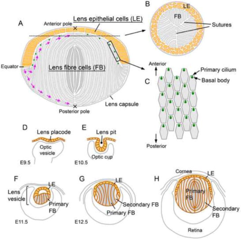
Fig. 1. Lens structure (A–C) and development (D–H).

(A) Most of the proliferative activity in the lens epithelium (yellow) is found in the region anterior to the equator. Cells that shift posterior to the equator exit the cell cycle and initiate terminal differentiation into lens fibre cells (grey). The apical and basal tips of the elongating fibres undergo directed migration (purple arrows) to the anterior and the posterior poles, respectively. At the poles, each fibre tip meets the tip from an equivalent fibre from another segment and collectively these aligned junctions form the lens sutures. The epithelial and fibre cells are contained within a thick extracellular matrix, the lens capsule. (B) A cross section around the anterior pole shows Y-shaped sutures formed by the aligned apical tips of the fibre cells. (C) A superficial view shows the hexagonal apical surfaces of the fibres with anteriorly polarised primary cilia/basal bodies. (D–H) Lens morphogenesis begins with the thickening of the surface ectoderm in the head region adjacent to the optic cup to form the lens placode (D). Following invagination to form the lens pit (E), the cells round up and separate from the surface ectoderm to form the lens vesicle (F). The cells in the posterior half of the lens vesicle elongate and undergo terminal differentiation to form the primary fibre cells that fill the vesicle lumen (G). The cells in the anterior half of the vesicle form an epithelial layer and this establishes the characteristic anterior-posterior polarity of the lens (G). Secondary fibre cells differentiate from the lens epithelial cells at the lens equator and progressively accumulate around the primary fibre cells so that the primary fibres are internalised and comprise the lens nucleus (H).
