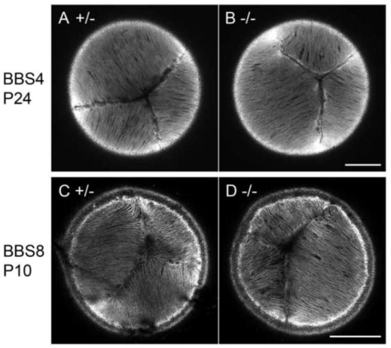
Fig. 4. Lens fibre cells maintain normal cellular alignment in BBS4 and BBS8 KOs.

Confocal sections of the anterior aspect of the lenses stained with phalloidin. In control lenses (A, C) apical tips of the fibres formed the typical Y-shaped suture pattern. This pattern was also detected in both BBS4 (B) and BBS8 (D) KOs. Scale bars 200 μm.
