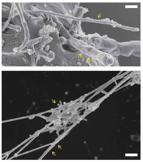
Figure 1.
Cellular and extracellular interactions of carbon nanotubes. The upper panel shows an SEM image of isolated MWCNTs (single arrow) or a bundle of MWCNTs (two arrows) entering human mesothelial cells. Reprinted from: Shi X, von dem Bussche A, Hurt RH, Kane AB, Gao H. Cell entry of one-dimensional nanomaterials occurs by tip recognition and rotation. Nat Nanotechnol. 2011;6(11):714–9, with permission from Nature Publishing Group. The lower panel shows a cluster of short-cut SWCNTs (single arrow) entrapped in chromatin fibers (two arrows) of purified neutrophil extracellular traps [see 159 for further details]. SEM courtesy of K. Hultenby, Karolinska Institutet.

