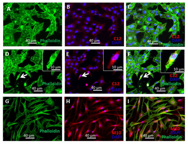
Figure 2. Intracellular localization of M10.
A–F, Lung fibroblasts were seeded on 4-chamber slides, serum-starved for 24 h followed by incubation for 24 h with M10 or scrambled peptide (10 μg/ml). Cells were fixed with 4% formaldehyde and stained with anti-Met (C12) antibody (A–F), or incubated with TAMRA-conjugated M10 (G–I). After labeling slides were mounted by Gold anti-fade reagent with Phalloidin and DAPI and visualized using a Leica DMI4000B fluorescence microscope equipped with Hamamatsu Camera Controller ORCA-ER.

