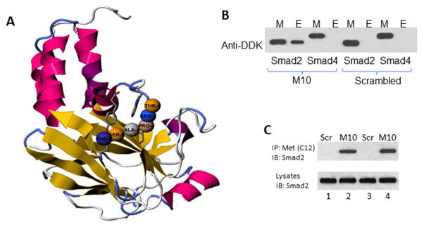
Figure 5. M10 and Smad2 interaction.
A, M10 in complex with Smad2. The interactive visualization of statistically significant (p < 0.02) binding of M10 (amino acids pro-3, ala-4, trp-7, glu-8, thr-9, and ser-10) with MH2 domain of Smad2. B, M10 interacts with Smad2 but not Smad4. DDK-tagged recombinant Smad2 and Smad4 were incubated with M10 as detailed in the Materials and Methods. Interacting complexes were captured with Protein G sepharose and subjected to immunoblotting with anti-DDK antibody. Peptide-protein interacting mixture (M) was used as a positive control. E stands for Eluted Interacting Complexes. C, Co-immunoprecipitation of M10 and Smad2 in scleroderma lung (lanes 1 and 2) and skin (lanes 3 and 4) fibroblasts. Cells were cultured in 100 mm plates to confluence, serum-starved overnight, incubated with M10 or scrambled peptide (Scr) for 24 h, collected, and subjected to immunoprecipitation as outlined in the experimental procedures.

