
Figure 2. Molecular Features of the Proband.
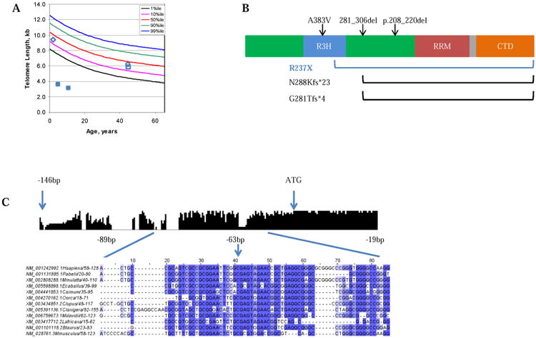
A) Lymphocyte telomere length, as measured by flow-FISH, for family NCI-165: proband, filled blue squares at age 5 and 13; brother, open blue diamond, father, open blue square, mother open blue circle. B) PARN contains three RNA-binding domains (the R3H domain [red], the RRM domain [RNA recognition motif, blue], two catalytic nuclease domains [green]), and a C-terminal domain (CTD, orange). A predicted bipartite NLS motif is located between the RRM domain and CTD (grey)27. Black arrows/brackets indicate previously reported PARN mutations associated with Hoyeraal-Hreidarsson syndrome 5, 14; The blue bracket indicates the deletion reported here. C) The histogram represents conservation amongst vertebrates across the PARN promoter region. Bases -89 to -19 (the region encompassing the 5′ UTR mutation detected in NCI-165-1) are shown in the detailed multiple sequence alignment below.