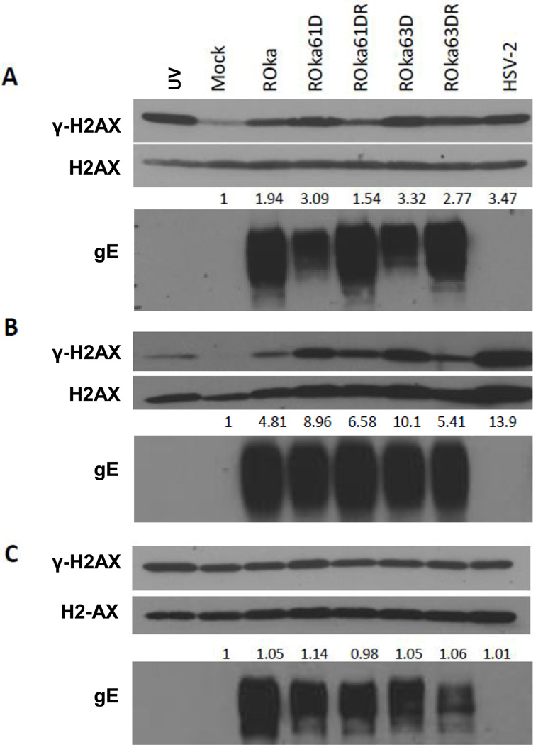
Fig. 1.
Immunoblot of γ-H2AX and H2AX expression in melanoma (MeWo) cells (A), human diploid fibroblasts (MRC-5 cells) (B) and U2OS osteosarcoma cells (C) infected with wild-type VZV, VZV mutants, and HSV-2. MeWo, MRC-5 and U2OS cells were infected with 1 × 10⁶ PFU of cell-associated VZV (ROka, ROka61D, ROka61DR, ROka63D, or ROka63DR) or with HSV-2 at an MOI of 0.5. At 48 hr after infection with VZV (or 24 hr after with HSV-2), the cells were lysed and equivalent amounts of cell lysates were immunoblotted with anti-γ-H2AX, anti-H2AX, and anti-VZV gE antibody. The numbers below the H2AX panels indicate the ratio of intensity of the γ-H2AX and H2AX bands in infected cell lysates divided by the ratio of the intensity of the γ-H2AX and H2AX bands in mock-infected cells. Densitometry was performed using Image J software. UV indicates cells that were treated to 50 mJ/cm2 of UV irradiation and 14 hr later lysates were prepared. The experiments were repeated and similar results were obtained.

