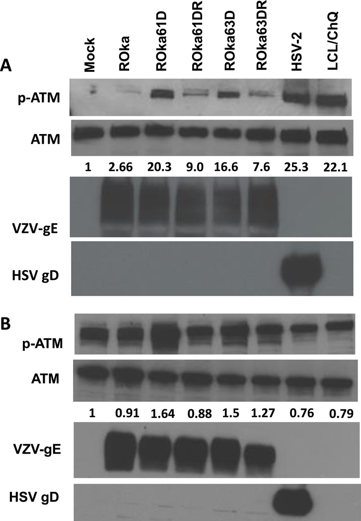
Fig. 4.
Immunoblot of phosphorylated-ATM and ATM expression in melanoma (MeWo) cells (A) and U2OS osteosarcoma cells (B) infected with wild-type or mutant VZV or HSV-2. Cells were infected with 1 × 10⁶ pfu of cell-associated VZV ROka, ROka61D, ROka61DR, ROka63D, ROka63DR, or infected with HSV-2 at an MOI of 0.5. At 48 hr after infection (or 24 hr after infection in HSV-2), the cells were lysed and equal amounts of clarified lysates were immunoblotted with anti-phosphorylated-ATM (Ser1981), ATM, VZV gE, and HSV gD. The numbers below the ATM panels indicate the ratio of intensity of the phosphorylated ATM and ATM bands in infected cell lysates divided by the ratio of the intensity of the phosphorylated-ATM and ATM bands in mock-infected cells. Densitometry was performed using Image J software. A chloroquine (ChQ) treated lymphoblastoid cell line (LCL) was a positive control for phosphorylation of ATM. The experiments were repeated with similar results.

