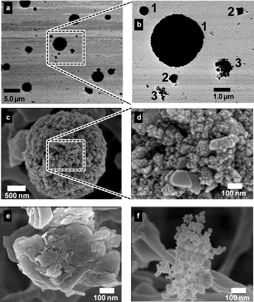
FIGURE 2.
Electron microscopy images from filter samples depict particle size and morphology. TEM images a and b show three types of particles of different shape and size: (1) b1, large spherically shaped particles; (2) b2, irregularly shaped particles; and (3) b3, smaller particle chains. SEM images c-f reveal that the larger spherical particles are actually composed of smaller nanoparticles 10–80 nm in size clustered into larger aggregates, c, d. Irregularly shaped particles have an amorphous structure, e. Chain agglomerates are composed of spherical nodules of 5–50 nm in size, f.

