
Fig. 4.
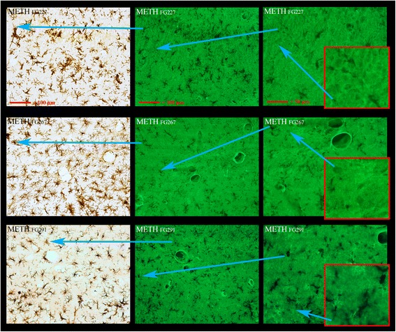
Pronounced microglia activation occurs with little or no apparent neurodegeneration. Dual labeling of microglia with IBA1 immunoreactivity and FJc to detect neurodegeneration is shown in the rostral and dorsal-medial area of the hippocampus. Pronounced microglial activation can be seen in the three left-hand panels, each for a different METH-treated animals (IDs present at the top left of panels). There was no evidence in this region in any of the three animals for terminal, axonal, or cell body labeling degeneration from FJc labeling in the remaining six panels. Again, as seen in the septum, the most intensely labeled structures were some of the vasculature present. The blue arrows show where the region of high magnification resides in the low magnification panels