
Fig. 7.
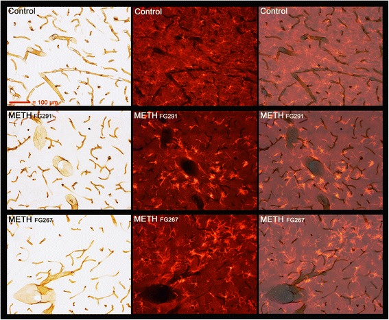
Activated microglia in the thalamus associated with vasculature after METH. Thalamic sections from a control and two METH-treated animals were double labeled using DAB labeling of an antibody to RECA1 and TRITC labeling of an antibody to IBA1. The far left panels show the DAB-labeled RECA1 immunoreactivity to vasculature through visible light and the middle panels show the TRITC-labeled IBA1 immunoreactivity to microglia through fluorescent illumination. The far right panels are a merger of the first two panels. The indigo arrows show regions of particular interest (see the “Results” section for details). All panels are of the same magnification