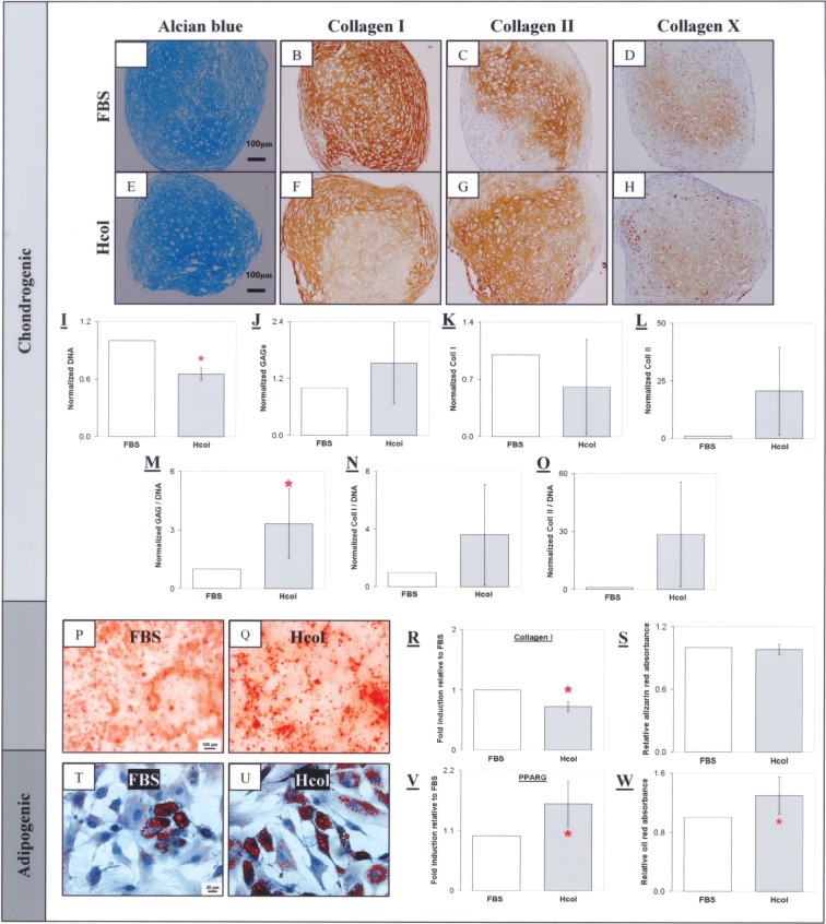
Figure 3.
Chondrogenic, osteogenic, and adipogenic differentiation of P3 MSCs. Chondrocytic lacunaes were found in the induced MSC pellets as indicated by the presence of glycoaminoglycans (GAGs) (A, E) and collagen II (C, G). Collagen I staining was stronger in the FBS pellet (B) compared to the Hcol group (F). A mild collagen X staining was found in both groups (D, H). Quantitative analysis indicated high cellularity in the FBS pellet (I) with lower total GAGs (J) and collagen II (L) compared to the serum free group. This was similarly observed with the DNA normalized values (M, O). There was enhanced GAGs and collagen II synthesis per cell maintained under the serum-free conditions. Conversely, the FBS group has a higher total collagen I (K) but the DNA normalized value was equivalent for both groups (N). The Hcol MSC exhibited enhanced chondrogenic differentiation compared to the FBS culture. Mineralization was observed in the FBS and Hcol groups with Alizarin red (P, Q). There was a lower collagen I gene expression in the Hcol culture (R) but calcification levels were equivalent in both groups (S). Lipid accumulation was observed in the cells under adipogenic induction as shown by oil red (T, U). However, adipogenesis was superior in the Hcol culture with higher PPARG gene expression (V) and higher lipid content (W). All controls stained negatively. *Significant difference between FBS and Hcol (p < 0.05, N = 4).

