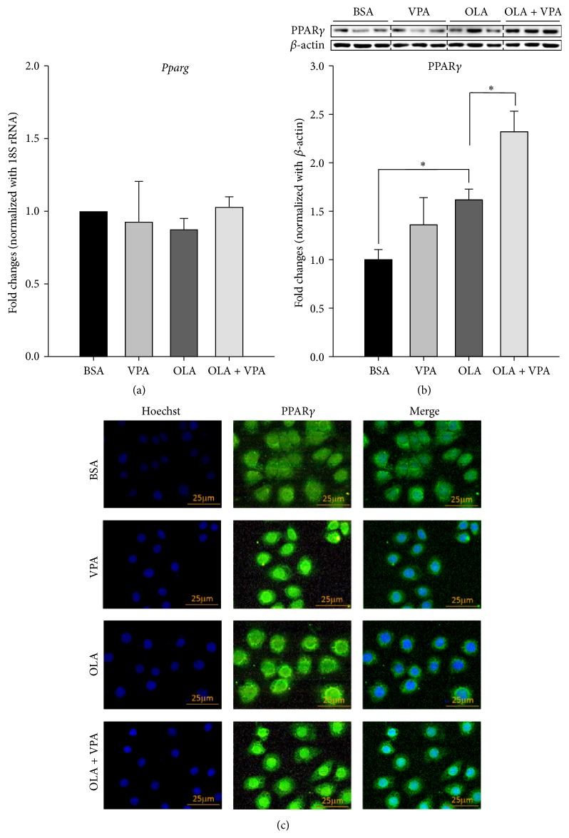
Figure 5.
VPA enhances oleic acids increased PPARγ protein expression and nuclear translocation, but not the mRNA levels. Real-time PCR (a), Western blotting (b), and immunofluorescence staining (c) were conducted following 24-hour treatment with BSA, VPA (1 mM), OLA (100 μM), or OLA plus VPA. For real-time PCR, relative fold changes were calculated using Ct values obtained from three independent experiments and are shown as means ± SEM. ∗ above the bars refer to significant differences (P < 0.05). Densitometric analyses for Western blotting were conducted for sample sets obtained from three independent experiments, and results are shown as means ± SEM. ∗ above the bars refer to significant differences (P < 0.05). In the immunofluorescence staining images, nuclear and intracellular PPARγ proteins were stained by Hoechst 33342- (blue) and DyLight 488-conjugated antibody (green), respectively.

