Figure 1.
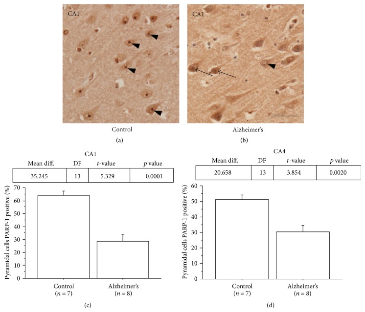
Nucleolar PARP-1 immunoreactivity in AD ranged from absent to dispersed and less intense compared to that of controls. ((a) and (b)) Representative immunostaining with diaminobenzidine (DAB) of human hippocampal pyramidal neurons in CA1 region. (a) Prominent nucleolar staining of PARP-1 (arrows) was seen in most of pyramidal neurons of a control case. (b) The nucleolar staining of PARP-1 ranged from absent (arrowheads) to a more dispersed pattern with less intensity of label (arrows) in pyramidal neurons of an AD case. ((c) and (d)) Percentages of CA1 and CA4 hippocampal pyramidal neurons with PARP-1 positive nucleoli were significantly lower in AD cases compared to controls. (Control, n = 8; AD, n = 8; ∗ p < 0.05.) Scale bar = 50 μm.

