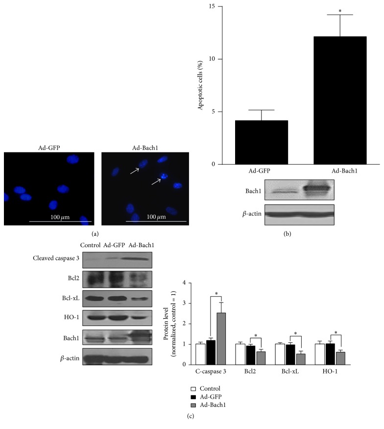
Figure 2.
Bach1 promotes apoptosis in cultured HMVECs. (a) HMVECs were infected with the adenoviruses (Ad-GFP or Ad-Bach1); then cells were fixed at 72 hours after infection, and nuclei were visualized by Hoechst 33342. Cells with nuclear condensation are indicated by white arrows. Bars, 100 μm. (b) Ad-GFP- and Ad-Bach1-infected HMVECs were seeded in 60 mm dishes, cultured for 72 hours, and then harvested and labeled with annexin V and PI. Cell apoptosis was quantified by identifying positively annexin V labeled cells via flow cytometry (n = 3; ∗ P < 0.05 versus Ad-GFP, upper panel). Bach1 protein levels were evaluated via Western blot (lower panel). (c) Cleaved caspase 3, Bcl2, Bcl-xL, HO-1, and Bach1 protein levels were determined via Western blot in HMVECs that had been infected with Ad-GFP or Ad-Bach1 (n = 3; ∗ P < 0.05 versus Ad-GFP).

