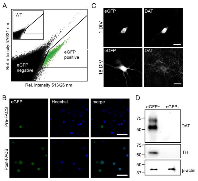
Figure 10. Fluorescence activated cell sorting and culturing of sorted postnatal dopaminergic neurons from Dat1-eGFP mice.
(A) FACS-purification of cells isolated from the midbrain of hemizygous Dat1-eGFP mice (P2–6). Representative scatter plot showing the emission intensity of sorted events at 513/26 nm against 576/21 nm after excitation at 488 nm. Events arise from sorting of a single cell suspension of ventral midbrain neurons from transgenic Dat1-eGFP mice. Insert shows a similar sorting from a WT animal. Note the appearance of a population of events at a higher 513/26 nm intensity for the Dat1-eGFP animal. These events are designated eGFP positive events (eGFP+), events outside this gate are designated eGFP negative events (eGFP−). (B) Confocal microscopy of cultured FACS sorted eGFP+ events at 1 DIV (upper panel) and 16 DIV (lower panel), showing eGFP reporter signal and DAT-ir. Sorted eGFP+ cells are viable and can be cultured for more than two weeks, in which period they will protrude extensions. Furthermore, expression of eGFP reporter signal and DAT-ir is detectable from 1 DIV. Scalebars = 50μm. (C) Visualization of FACS-purified cells from the Dat1-eGFP midbrain suspensions confirmed eGFP+ phenotype. Upper panels; epifluorescence microscopy of cells before FACS (pre-FACS), Lower panels; epi-fluorescence microscopy of FACS-purified eGFP+ cells (post-FACS). Hoechst, a nuclear staining marker, was included to discriminate between viable and non-viable cells during the sorting process. Scalebars = 50μm. (D) Immunoblotting confirms a dopaminergic phenotype of the eGFP+ sorted cell population. Representative immunoblots demonstrate enrichment of the dopaminergic markers DAT and TH in cell lysates from the eGFP+ cell population while DA markers are completely absent in the GFP− population (n = 3). Cell lysates from purified eGFP+ and eGFP− cells were analyzed with SDS-PAGE followed by immunoblotting for anti-DAT and anti-TH antibody as described in Material and Methods. DAT and TH is exclusively expressed in lysates from eGFP+ sorted cells. Note that anti-DAT antibody recognizes two bands corresponding to the mature (~70 kDa) and immature (~55 kDa) isoforms of DAT while anti-TH recognizes one band at (~55 kDa). β-actin was used to verify equal loading for respective cell population.

