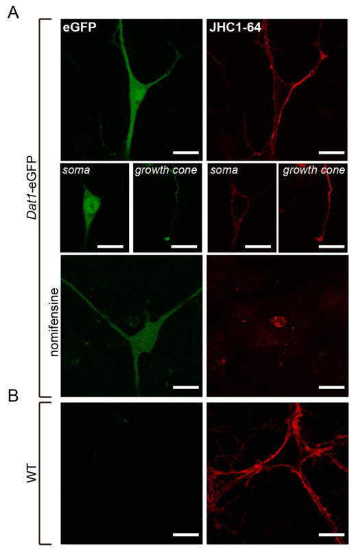
Figure 8. The fluorescent cocaine analogue JHC 1–64 labels eGFP-expressing cells in live midbrain dopaminergic cultures from Dat1-eGFP mice.
(A) Confocal images of cultured midbrain neurons from Dat1-eGFP mice labeled with JHC 1–64. The representative images show the signal corresponding to the somatodendritic compartment, and growth cones (8A, middle panels). Incubation with the DAT specific antagonist nomifensine (100 μM) prior to and during JHC 1–64 incubation completely abolished the binding. The JHC 1–64 signal distributes to the plasma membrane of the somas and to the neuronal extensions as well as a clear signal is found corresponding to the growth cones. Importantly, JHC 1–64 exclusively labeled neurons from Dat1-eGFP mice that also displayed an eGFP signal. The eGFP signal was homogenously distributed in the entire cytoplasm with intense expression in the somatic region. (B) Confocal images of cultured midbrain neurons from non-transgenic WT mice labeled with JHC 1–64. Despite JHC 1–64 labeling of several neurons, we observe no eGFP signal. Scale bar = 20 μm. Images shown are representative of at least three independent experiments.

