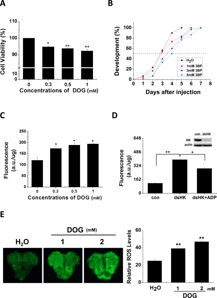
Figure 2. Inhibitor effects on cell viability, developmental delay, and ROS activity.
(A) Cell viability changes. HzAm1 cells were treated with various concentrations of DOG for 48 h, and viable cells were measured using the MTT assay; the values represent the mean ± S.D. of at three independent experiments. (B) Developmental delay caused by 3BP injection. Day 1 nondiapause-destined pupae were injected with 3 μl of 3BP at various concentrations (1 mM 3BP, n = 30; 3 mM 3BP, n = 30; and 5 mM 3BP, n = 30), and pupal stemmata were examined as a marker for pupal development. As a control, pupae were also injected with 3 μl of H2O (n = 30). (C) Effects of DOG on ROS activity. HzAm1 cells were treated with DOG for 24 h and fluorescence was measured. (D) ROS activity changes due to Har-HK knockdown. HzAm1 cells were transfected with dsGFP (con) or dsHK for 36 h and then treated with 1 mM ADP for 12 h; each point represents the mean ± S.D. of three independent replicates. (E) Effects of DOG on brain ROS activity. Day 1 nondiapause-destined pupae were injected with 3 μl of DOG (n = 5), and fluorescence was then measured. The * denotes p < 0.05, and ** denotes p < 0.01 as determined by the independent t-test.

