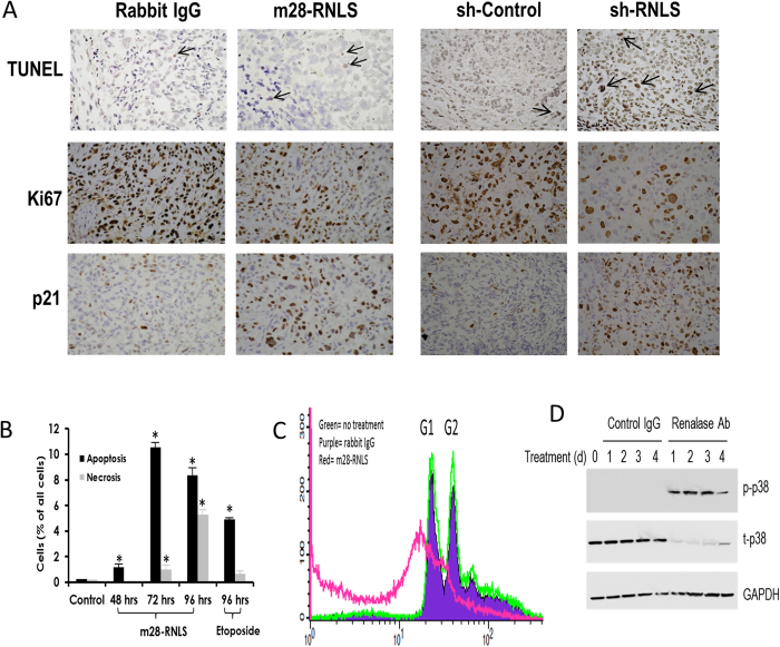
Figure 3. Inhibition of RNLS signaling induces apoptosis and cell cycle arrest.
(A) Representative images of sections from BxPC3 xenografted tumors (n = 14 each) treated with anti-m28-RNLS or control rabbit IgG, and of xenografts of Panc1 cells transduced with RNLS shRNA (sh-RNLS) or control (sh-Control). Tissues are stained for TUNEL (arrows: positive cells), cell proliferation marker Ki67 (brownish stain), and cell cycle inhibitor p21 (brownish stain). (B) FACS analysis of Panc1 cells in culture treated with either m28-RNLS (30 μg/ml) or 100 μM etoposide (positive control) for 4 days; n = 3, *indicates p < 0.05. (C) Effect of m28-RNLS on cell cycle of Panc1 cells determined by FACS analysis; green curve: no treatment, purple curve: rabbit IgG, red curve: m28-RNLS 30 μg/ml. (D) Panc1 cells treated with polyclonal ab31291 or with goat IgG as a negative control, and cell lysates probed for activation of p38 by western blot from day 0 to 4. Representative blot, n = 4.

