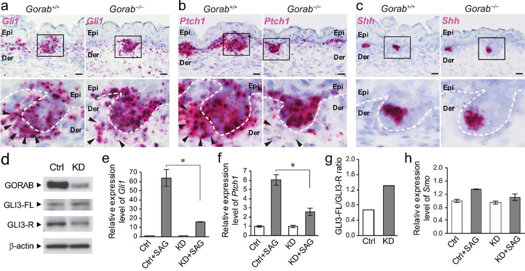
Figure 4. Hh signaling pathways is impaired in mutant mesenchymal cells.
(a–c) Expression of Gli1, Ptch1, and Shh in E15.5 skins of control (Gorab+/+) and homozygous Gorab mutants (Gorab−/−) by in situ hybridization. Dotted lines illustrate the basement membrane. Arrowheads point to dermal papilla cells. n=4. (d) Expression of GORAB, full-length GLI3 (GLI3-FL, ≈190 kDa), and repressor form of GLI3 (GLI3-R, ≈85 kDa) in Gorab knockdown (KD) mouse embryonic fibroblasts (MEFs) by western blotting. (e and f) Quantification of Gli1 and Ptch1 mRNA by quantitative RT-PCR after SAG treatment in control (Ctrl) and KD MEFs. Asterisk (*) indicates p < 0.01. (g) Quantification GLI3-FL/GLI3-R ratio in control and KD MEFs as shown in a. (h) Quantification of Smo mRNA by quantitative RT-PCR in SAG-treated control and KD MEFs. All experiments were performed three times. Scale bars: 20 µm.

