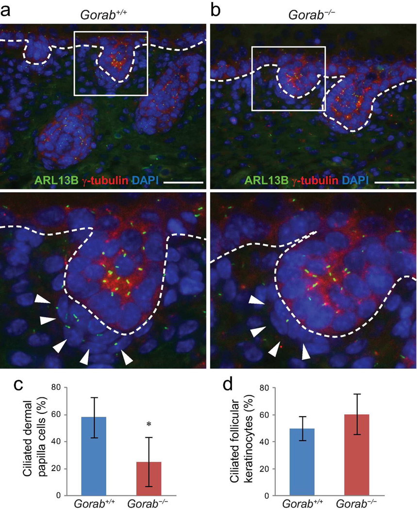
Figure 5. Formation of primary cilia is impaired in dermal condensate cells of Gorab mutants.
(a) Immunofluorescence labeling of primary cilia with ARL13B (green) in dorsal skin of E18.5 control (Gorab+/+) and homozygous Gorab mutants (Gorab−/−). Basal bodies labeled with γ-tubulin (red); nuclei were stained with DAPI (blue). Lower panels are magnified boxed hair germs in the upper panels. Dotted lines illustrate the basement membrane. Arrowheads point to dermal papilla cells. (c and d) Quantification of ciliated dermal condensate cells (c) and follicular keratinocytes (d). A minimum of three animals were evaluated for each genotype. Asterisk (*) indicates p<0.05. Scale bars: 50 µm.

