Figure 7.
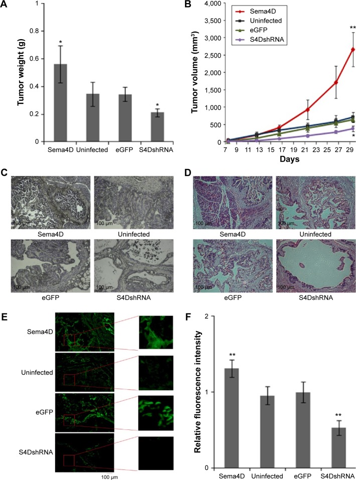
Semaphorin 4D (Sema4D) promotes tumor growth and vascularity in Caco-2 cells with high vascular endothelial growth factor background.
Notes: Caco-2 cells were treated the same as HCT-116, and grouped into four groups: uninfected, enhanced green fluorescent protein (eGFP) control, Sema4D coding, and Sema4D short hairpin RNA (shRNA) group. Nude mice were injected with cells of four groups respectively; as a result, tumors were harvested and detected as well. (A) The results of tumor weights harvested in four groups of Caco-2 cells are shown in the bar graph (n=5; *P<0.05, relative to both uninfected and eGFP group, PSema4D =0.01, PS4DshRNA =0.01). Bars indicate mean of five tumor weights ± standard error. (B) Tumor growth curve for the four groups of Caco-2 cells, tumor volume measurement in mm3 are shown (n=5; *P<0.05, **P<0.01, relative to both uninfected and eGFP groups). Bars indicate mean of five tumor volume ± standard error. (C) Immunohistochemical analysis of Sema4D in tumor tissues harvested from four indicated groups. The result shows significant differences within the four indicated groups. (D) The result of hematoxylin and eosin staining for tissues from different groups. (E) CD31 immunofluorescence staining of frozen section of tumors derived from four indicated groups, for evaluation of vascular density. (F) Bar graphs showed the results of measurement of vascular content from indicated groups, which are determined by the average vessel density in ten CD31-stained sections from each group (n=10; **P<0.01, relative to eGFP group, PSema4D =1.4E-05, PS4DshRNA =5.3E-08). Bars indicate mean of vessel density in ten CD31-stained sections ± standard error.

