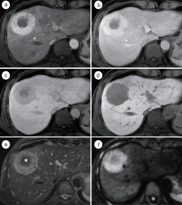
Fig. 2.
Mass-forming type ICC which mimics the enhancement pattern of HCC on Gd-EOB-MRI in a 49-year-old male with alcoholic liver disease. A round mass which is located in segment 4 and 8 of the liver (a) shows arterial enhancement in the peripheral portion of the mass. The arterially-enhancing portion of the mass shows persistent enhancement on the PVP (b), but hypointensity on the TP (c) and HBP (d). Note the progressive enhancement of the central portion of the mass (a-c). On T2-weighted image, the peripheral portion of the mass (e) shows hyperintensity whereas the central portion (*) shows hypointensity due to the abundant fibrous stroma. On diffusion-weighted image (b value=800 sec/mm2), the mass (f) demonstrates a target appearance. Histopathologic examination confirmed this tumor as cholangiocarcinoma.

