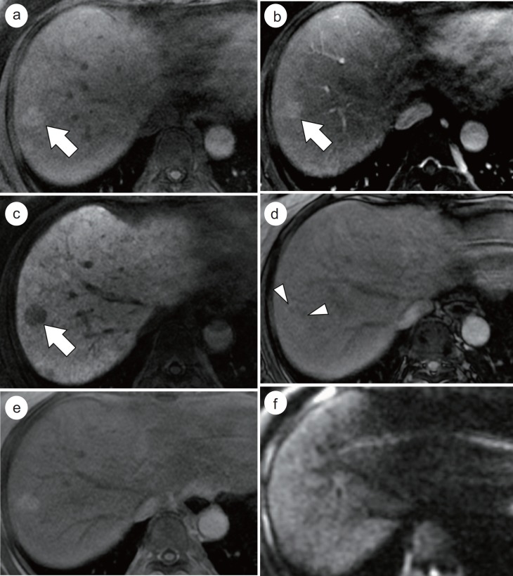
Fig. 4.
Histopathologically-confirmed DN in a 55-year-old female with liver cirrhosis. A 2 cm nodule (arrows) in segment 8 of the liver shows hyperintensity on precontrast T1-weighted image (a), and no definite enhancement on the HAP image (b). On the HBP imaging (c), the nodule demonstrates hypointensity. On opposed-phase imaging (d), there are foci with decreased signal intensity compared with in-phase imaging (e), which suggest intratumoral fat component. On diffusion-weighted image (b value=800 sec/mm2), the nodule (f) shows isointensity.

