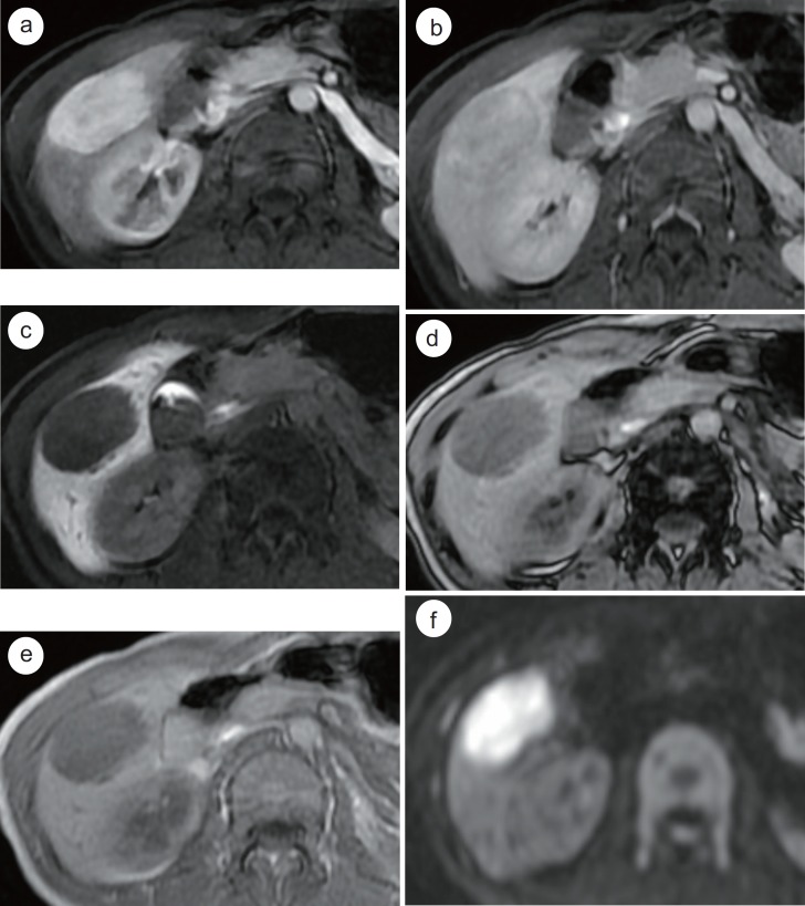
Fig. 7.
Histopathologically-confirmed hepatic AML in a 33-year-old female without chronic liver disease. On Gd-EOB-MRI, a 5 cm oval mass in segment 5 of the liver shows homogeneous arterial enhancement (a), iso- to hypointensity on the PVP (b), and homogeneous hypointensity on the HBP images (c). No signal drop is noted in opposed-phase imaging (d) compared with in-phase imaging (e). Note the diffusion restriction of the hepatic mass (f) (DWI with b value of 800 sec/mm2).

