

Abstract
Context:
The infrapatellar fat pad (IFP) is in the anterior knee compartment and may be a major pain generator.
Evidence Acquisition:
A PubMed database search using the terms Hoffas fat pad, anterior interval, and infrapatellar fat pad was performed from the years 1970 to 2015.
Study Design:
Clinical review.
Level of Evidence:
Level 5.
Results:
Limited research exists examining the role of the IFP in relation to potential treatment and rehabilitation implications.
Conclusions:
Alterations in IFP mobility, whether the result of postsurgical scarring or faulty movement patterns, result in pain and disability in a variety of patient populations. The majority of treatment approaches are driven by the surgical technique.
Keywords: infrapatellar fat pad, Hoffas fat pad, anterior knee pain
Anatomy, Vascularization, Innervation
The infrapatellar fat pad (IFP) is in the anterior knee compartment and may be a major pain generator.21 The exact role of the IFP remains unknown, but the IFP retracts into the joint posteriorly during flexion and moves anteriorly away from the tibia during extension, indicating that IFP mobility within the anterior interval may be crucial.8 If fibrosis or scarring of the IFP occurs, it can lead to adhesions in the anterior interval, which can decrease excursion of the patellar tendon.11 Additionally, the IFP is capable of eliciting high levels of pain.10 Dye et al10 probed structures of the human knee without the use of intra-articular anesthesia and determined that the IFP was an extremely sensitive structure capable of producing severe pain. During this nonanesthetized probing of the IFP, the pain elicited was so severe that the study was nearly stopped.10
The IFP has space-filling properties, and in the presence of trauma, it may expand and act as a pain generator.11 An inflamed IFP can bulge on either side of the patellar tendon, with the synovial membrane being compressed against the femoral condyles. This compression of the synovial membrane can cause knee effusions and potentially lead to pain.11
The IFP is richly vascularized by an anastomotic network (Figure 1).13 The center of the IFP has low vascularization and is therefore regarded as a suitable entry point in the case of arthroscopic surgery to limit bleeding and damage.13
Figure 1.

Arterial supply to the fat pad: (a) medial superior genicular artery; (b) descending genicular artery; (c) medial superior genicular artery, additional branch; (d) lateral superior genicular artery; (e) lateral inferior genicular artery; (f) arterial tibial recurrent artery; and (g) medial inferior genicular artery. Reprinted with permission from Kohn et al.13
Witonski et al24 observed synovitis and swelling of the fat pad in knees after anterior cruciate ligament (ACL) rupture in 20 patients (6 male, 14 female; mean age, 18.6 years), as well as an increased prevalence of substance-P fibers in individuals with anterior knee pain. Substance P increases sensitivity to nociceptive signals and promotes inflammation, which can have a direct influence on pain. In previously reported cases of anterior knee pain in 20 women (mean age, 18.7 years), neuropeptide-containing fibers were significantly more common in the medial retinaculum (P < 0.005) and fat pad (P < 0.001) compared with groups without anterior knee pain.24 High rates of substance-P fibers are found around vessels of the IFP, and as such, can cause vasodilation and migration of white blood cells from capillaries to surrounding tissue, resulting in IFP edema. This edema can lead to IFP impingement in the knee joint as well as ischemia and lipomatous tissue necrosis.3
The IFP is a possible source of pain in the anterior knee with innervation of branches of the posterior tibial nerve.21 Additionally, numerous nerves innervating other structures in the knee also have branches to the IFP.12 These include nerves in the vastus lateralis and medialis, a terminal branch of the obturator nerve, the saphenous nerve, and the common peroneal nerve.12 The health and integrity of the fat pad is also crucial for other reasons. The adipose tissue of the IFP in the knee serves as an abundant source of mesenchymal stem cells, which have the ability to differentiate into multiple different types of tissue, including cartilage and bone.6 Under appropriate in vitro conditions, these cells can differentiate into chondrocytes, which could be an important clinical function for cartilage repair and replacement techniques.7
Infrapatellar Fat Pad Biomechanics
The exact role of the IFP is not completely understood. Some researchers have hypothesized that it has a biomechanical role, while others suggest it is a reservoir for reparative cells.2,4,15,22 From a biomechanical perspective, the IFP may enhance gliding between the femoral condyles and joint capsule.20,21 Knee mechanics are altered when IFP adhesions occur.4
The IFP is mobile and changes shape, position, pressure, and volume as the knee moves through its full range of motion.2,5,7,8 During flexion, the angle between the patellar tendon and the anterior border of the tibia decreases, displacing the fat pad posteriorly.5,8 During knee extension, the IFP moves away from the anterior tibia.5,7,8 With postoperative IFP scarring, normal mechanics may be compromised. Cadaveric research demonstrates that IFP adhesions may create a patellar infera.1 This change in patellar position and patellar tendon adhesion alters the effectiveness of the extensor mechanism, decreasing the effective moment arm and requiring a greater quadriceps force to produce the same knee extension force (Figure 2).1
Figure 2.

Infrapatellar fat pad biomechanics. PFJR, patellofemoral joint reaction force; PT, patellar tendon. Reprinted with permission from Ahmad et al.1
During knee flexion, the patella normally engages the trochlear groove and translates distally and medially. When IFP adhesions are present, the patella may engage the groove prematurely and result in medial tracking.1 A similar effect is seen during knee extension as the relatively shorter patellar tendon length (secondary to the adhesions) decreases patellar mobility and creates resistance to lateral translation at full extension.1
Infrapatellar fat pad adhesions can alter tibial biomechanics changes as well. The shortened patellar tendon can direct an anterior force on the tibia, increasing tibial translation between 30° and 60° of flexion.1 These adhesions caused the contact areas on the medial and lateral tibial plateau to shift posteriorly.1,23
In addition to kinematic changes, IFP adhesions can alter knee contact forces.2 In a normal knee, the IFP pressures significantly increase at flexion angles greater than 100° and extension angles less than 20°.2 The volume of the anterior interval is directly related to pressure throughout the knee range of motion.2 An edematous fat pad can irritate surrounding tissues.2
Clinical Assessment and Treatment of Surgical and Nonsurgical Patients
Surgical treatment of pathology within the anterior interval of the knee is best performed using an arthroscopic approach with insufflation of the knee.21 This may be accomplished by injection of the joint with 120 to 180 mL normal saline or lactated ringers solution with an 18-gauge needle via a superolateral approach. Distension of the joint enables the surgeon to identify appropriate portal locations, protects intra-articular structures while inserting surgical instruments, and increases intra-articular pressure, which may minimize bleeding.21
An anterolateral portal located several millimeters proximal to the inferior pole of the patella may allow easier visualization of the anterior interval structures21 with either the 30° or 70° arthroscope. A second portal slightly proximal and medial to the medial margin of the IFP may allow better instrumentation of the anterior interval.21
The most common finding within the anterior interval is scarring of the IFP.21 With significant scarring, the intermeniscal ligament may be scarred to the anterior cortex of the tibia (Figure 3). A subtle finding is often decreased excursion of the anterior third of the medial meniscus with flexion beyond 60°.
Figure 3.
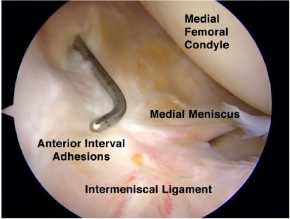
Intermeniscal ligament scarring.
The goal of surgery for fibrosis and scarring of the IFP and anterior interval is to lyse or ablate the fibrotic tissue and to preserve as much normal IFP tissue as possible (Figure 4, a and b).1,21 In the face of space occupying lesions of the anterior interval, such as cystic lesions, only the abnormal or hypertrophic tissue should be removed. Removing normal IFP tissue may create excessive postoperative swelling, bleeding, and pain and may inhibit the postoperative knee range of motion and rehabilitation, creating a scenario of more scarring within the anterior interval. Both the surgeon and therapist must be aware of this potential.
Figure 4.

Surgical fat pad release.
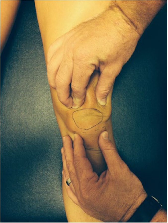
Fat pad restrictions are common following knee surgery but can be seen in nonsurgical patients with anterior knee pain as well.7 IFP restrictions can be assessed on physical examination by moving the IFP medially and laterally, determining restriction of motion compared with the contralateral and uninjured lower extremity (Figure 5, a and b).The IFP may be tender and/or firm.9,14,18 Pain elicited by direct pressure (Hoffa test) may indicate fat pad restrictions.17,21 Additionally, the superior glide and superior tipping of the patella is compared for motion restrictions (Figure 6)14,19,21 as the involved knee is flexed to approximately 60°. The inferior pole of the patella should tip upward, pulling the patellar tendon away from the anterior interval space (Figure 7, a and b). Another technique of assessing anterior interval adhesion is by passively hyperextending the patient’s knee in a supine position (Figure 8).14,16 In the hyperextended position, the patient should feel a stretch in the hamstring and posterior knee if the anterior interval allows the fat pad to glide anteriorly. If the patient experiences pain, pinching, or pressure in the anterior interval, there may be IFP restriction.
Figure 5.

(a) Medial and (b) lateral fat pad glide.
Figure 6.
Superior patella glide.
Figure 7.
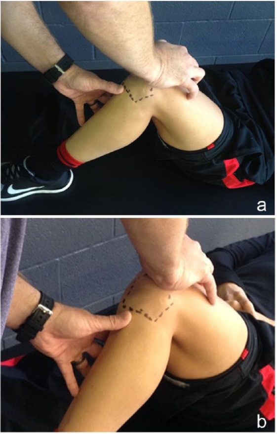
(a) Patellar tilt. (b) Rear view.
Figure 8.
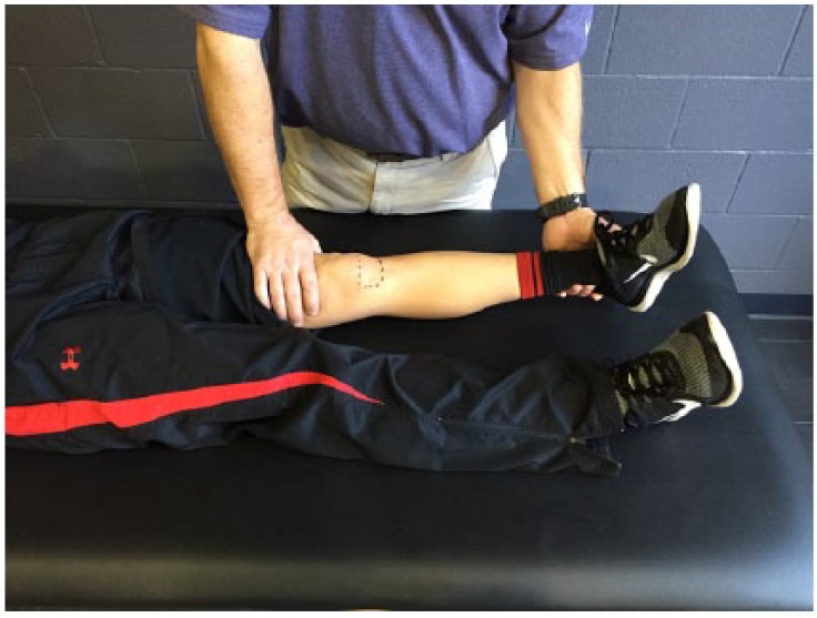
Passive terminal knee extension.
Patients with IFP adhesions often complain of anterior knee pain during flexion and extension of the knee joint.17,21 Squatting, stair climbing, jumping, and running tend to aggravate these symptoms.17,21 Quadriceps length (tightness) should be assessed; stretching the quadriceps and anterior hip structures may improve symptoms of IFP restrictions.7
Conclusion
Infrapatellar fat pad pathophysiology from postsurgical scarring or faulty movement patterns may result in pain and disability in a variety of patient populations.

Footnotes
The authors report no potential conflicts of interest in the development and publication of this article.
References
- 1. Ahmad CS, Kwak SD, Ateshian GA, Warden WH, Steadman JR, Mow VC. Effects of patellar tendon adhesion to the anterior tibia on knee mechanics. Am J Sports Med. 1998;26:715-724. [DOI] [PubMed] [Google Scholar]
- 2. Bohnsack M, Hurschler C, Demirtas T. Infrapatellar fat pad pressure and volume changes of the anterior compartment during knee motion: possible clinical consequences to the anterior knee pain syndrome. Knee Surg Sports Traumatol Arthrosc. 2005;13:135-141. [DOI] [PubMed] [Google Scholar]
- 3. Bohnsack M, Meier F, Walter GF, Hurschler C, Schmolke S, Wirth CJ. Distribution of substance-P nerves inside the infrapatellar fat pad and the adjacent synovial tissue: a neurohistological approach to anterior knee pain syndrome. Arch Orthop Trauma Surg. 2005;125:592-597. [DOI] [PubMed] [Google Scholar]
- 4. Bohnsack M, Wilham A, Hurschler C. Biomechanical and kinematic influences of a total infrapatellar fat pad resection of the knee. Am J Sports Med. 2004;32:1873-1880. [DOI] [PubMed] [Google Scholar]
- 5. Donell ST. The synovial folds of the patellofemoral joint: a dynamic study. Clin Anat. 1992;5:107-112. [Google Scholar]
- 6. Dragoo J, Samimi B, Zhu M, et al. Tissue-engineered cartilage and bone using stem cells from human infrapatellar fat pads. J Bone Joint Surg Br. 2003;85:740-747. [PubMed] [Google Scholar]
- 7. Dragoo JL, Johnson C, McConnell J. Evaluation and treatment of disorders of the infrapatellar fat pad. Sports Med. 2012;42:51-67. [DOI] [PubMed] [Google Scholar]
- 8. Dragoo JL, Phillips C, Schmidt JD. Mechanics of the anterior interval of the knee using open dynamic MRI. Clin Biomech. 2010;25:433-437. [DOI] [PubMed] [Google Scholar]
- 9. Duri ZA, Aichroth PM, Dowd G, Ware H. The fat pad and its relationship to anterior knee pain. Knee. 1997;4:227-236. [Google Scholar]
- 10. Dye SF, Vaupel GL, Dye CC. Conscious neurosensory mapping of the internal structures of the human knee without intra-articular anesthesia. Am J Sports Med. 1998;26:773-777. [DOI] [PubMed] [Google Scholar]
- 11. Gallagher J, Tierney P, Murray P, O’Brien M. The infrapatellar fat pad: anatomy and clinical correlations. Knee Surg Sports Traumatol Athrosc. 2005;13:268-272. [DOI] [PubMed] [Google Scholar]
- 12. Kennedy JC, Alexander IJ, Hayes KC. Nerve supply of the human knee and its functional importance. Am J Sports Med. 1982;10:329-335. [DOI] [PubMed] [Google Scholar]
- 13. Kohn D, Deiler S, Rudert M. Arterial blood supply of the infrapatellar fat pad. Arch Orthop Trauma Surg. 1995;114:72-75. [DOI] [PubMed] [Google Scholar]
- 14. Kumar D, Alvand A, Beacon J. Impingement of infrapatellar fat pad (Hoffa’s disease): results of high-portal arthroscopic resection. Arthroscopy. 2007;23:1180-1186.e1. [DOI] [PubMed] [Google Scholar]
- 15. Lemon M, Packham I, Narang K. Patellar tendon length after knee arthroplasty with and without preservation of the infrapatellar fat pad. J Arthroplasty. 2007;22:574-580. [DOI] [PubMed] [Google Scholar]
- 16. McConnell J. Fat pad irritation: a mistaken patellar tendonitis. Sport Health. 1992;9(4):7-9. [Google Scholar]
- 17. Metheny J, Mayor M. Hoffa disease: chronic impingement of the infrapatellar fat pad. Am J Knee Surg. 1988;1:134-139. [Google Scholar]
- 18. Paulos L, Swanson SC, Stoddard GJ, Barber-Westin S. Surgical correction of limb malalignment for instability of the patella: a comparison of 2 techniques. Am J Sports Med. 2009;37:1288-1300. [DOI] [PubMed] [Google Scholar]
- 19. Richmond JC, Al Assal M. Arthroscopic management of arthrofibrosis of the knee, including infrapatellar contraction syndrome. Arthroscopy. 1991;7:144-147. [DOI] [PubMed] [Google Scholar]
- 20. Smillie IS. Diseases of the Knee Joint. London, England: Churchill Livingstone; 1974. [Google Scholar]
- 21. Steadman JR, Dragoo JL, Hines SL, Briggs KK. Arthroscopic release for symptomatic scarring of the anterior interval of the knee. Am J Sports Med. 2008;36:1763-1769. [DOI] [PubMed] [Google Scholar]
- 22. Tanaka N, Sakahashi Sato E. Influence of the infrapatellar fat pad resection in a synovectomy during total knee arthroplasty in patients with rheumatoid arthritis. J Arthroplasty. 2003;18:897-902. [DOI] [PubMed] [Google Scholar]
- 23. van Eijden TM, Kouwenhoven E, Weijs WA. Mechanics of the patellar articulation: effects of patellar ligament length studied with a mathematical model. Acta Orthop Scand. 1987;58:560-566. [DOI] [PubMed] [Google Scholar]
- 24. Witonski D, Wagrowska-Danielewicz M. Distribution of substance-P nerve fibers in the knee joint in patients with anterior knee pain syndrome. Knee Surg Sports Traumatol Arthrosc. 1999;7:177-183. [DOI] [PubMed] [Google Scholar]