
Figure 1.
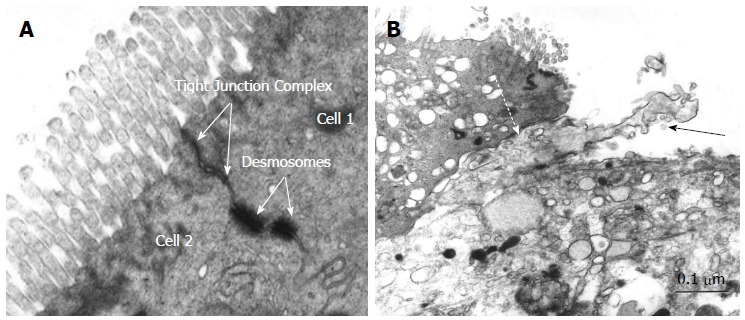
Electron microscopy of pouch epithelium tight junction complex in non-inflamed pouch (A) and electron micrograph demonstrating dendritic cell penetrating between two epithelial cells in pouchitis (B). Bar = 0.1 μm. Solid arrow: Dendritic cell; Dashed arrow: Tight junction complex between epithelial cell and dendritic cell.