
Figure 2.
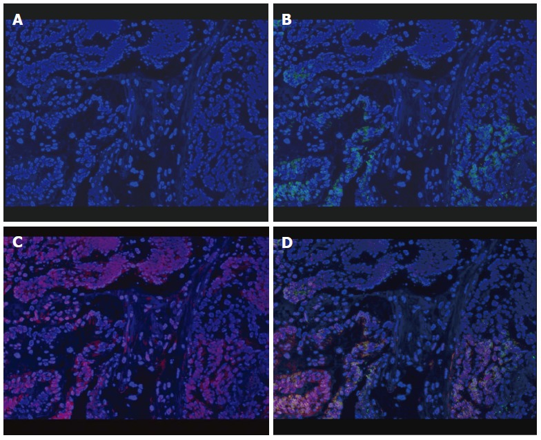
Enrichment of Fusobacterium nucleatum in colorectal cancer detected by FISH. Graph A: Colorectal cancer (CRC) specimen stained with DAPI. Cell nuclei were stained in blue; B: CRC specimen stained with both DAPI and universal bacterial probe (EUB338). Bacterial conserved regions were stained in green; C: CRC specimen stained with both DAPI and Fusobacterium specific probe (FUSO). The Fusobacterium nucleatum (F. nucleatum) specific regions were stained in red; D: CRC specimen triply stained with DAPI, EUSO and EUB338. Cell nuclei (in blue), bacterial conserved regions (in green) and F. nucleatum specific regions (in red) were clearly visible (all × 400).