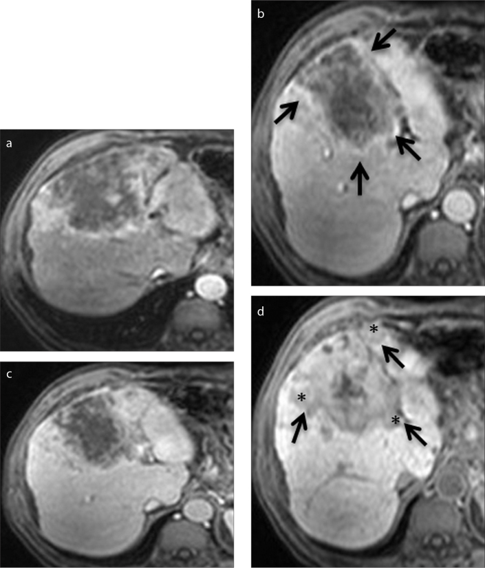
Figure 3.
a–d. A 52-year-old man with cirrhotic liver. Axial contrast-enhanced arterial (a), portal venous (b), delayed (c), and hepatobiliary (d) phase Gd-BOPTA-enhanced MRI scans show a heterogeneous mass that is histopathologically proven to be HCC. Capsule is best seen on the portal venous phase image (b, arrows), whereas tumor margin is best visualized on hepatobiliary phase images (d, arrows). Capsular disruption and irregular tumor margin are present (d, asterisks).

