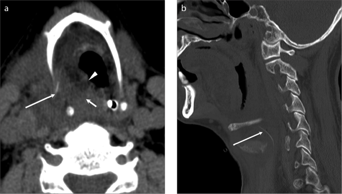
Figure 1.
a, b. Ingested fish bone with perforation of the right hypopharynx. Axial (a) and sagittal (b) contrast-enhanced CT images of the neck demonstrate a linear hyperdensity lying in the right extra laryngeal space (long arrows) suggestive of ingested fish bone. There is significant edema in the right paralaryngeal area (a, short arrow) resulting in mild narrowing of the airway (a, arrowhead).

