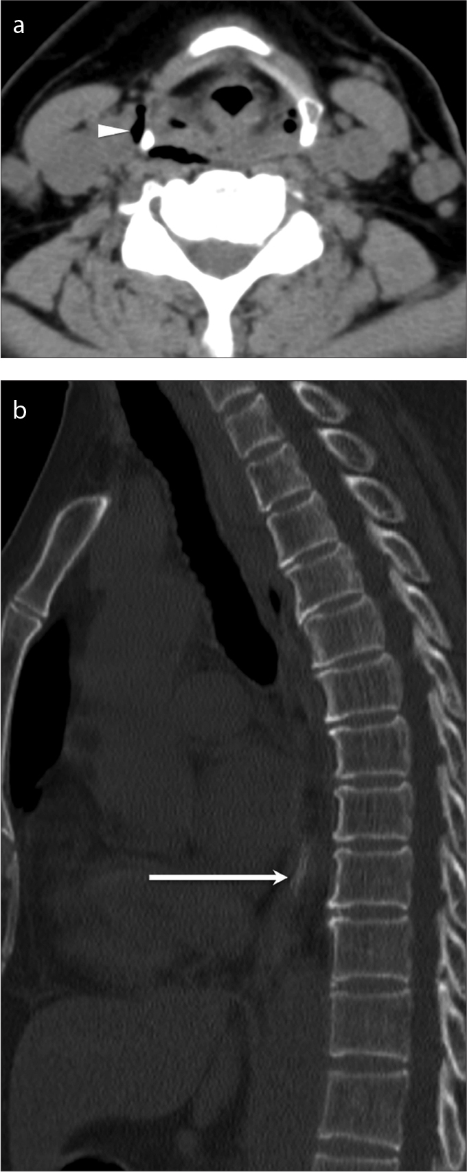
Figure 6.

a, b. Ingested fish bone with perforation of the upper esophagus, and air in the deep spaces of the neck. Axial unenhanced CT of the neck (a) and sagittal unenhanced CT of the thorax (b) demonstrate a linear hyperdensity (b, arrow) in the distal esophagus suggestive of an ingested fish bone causing perforation of the upper esophagus resulting in air in the deep spaces of the neck (a, arrowhead). The perforation of upper esophagus was confirmed by esophagoscopy; the fish bone itself had moved into distal esophagus as seen on CT.
