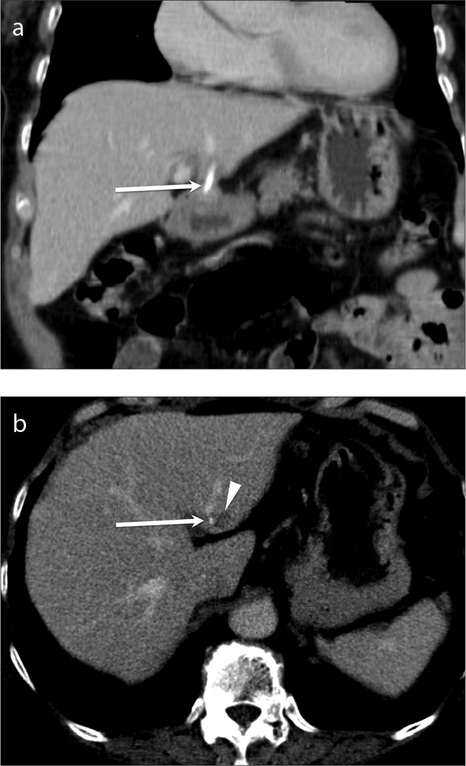
Figure 8.

a, b. Ingested fish bone migrating into the liver from perforation of the stomach resulting in a hepatic abscess. Contrast-enhanced coronal (a) and axial (b) CT of the abdomen demonstrates a linear hyperdensity suggestive of ingested fish bone (arrows) within the liver parenchyma with small surrounding hypodensity suggestive of developing abscess (b, arrowhead).
