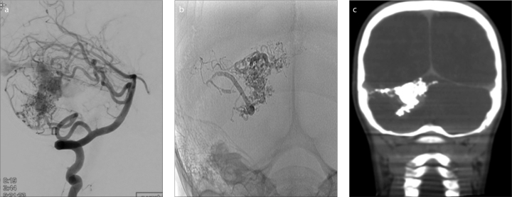
Figure 3.
a–c. A five-year-old male with a ruptured cerebellar AVM. Lateral subtracted-DSA (a) prior to embolization shows the AVM fed by posterior inferior cerebellar artery and superior cerebellar artery (SCA) branches. Postembolization AP plain radiography (b) and coronal XperCT (c) show partial PHIL embolization of AVM via SCA branches with minimal artifact noted on CT.

