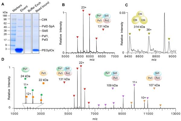
Figure 3.
Affinity isolation, peptide elution and native MS analysis of the endogenous GINS assembly from budding yeast. (A) SDS-PAGE separation and Coomassie staining to assess the post-elution sample handling steps. Elution was performed with 2 mM PEGylOx, which was later removed by buffer exchange into 150 mM ammonium acetate, 0.01 % Tween-20. (B) The native MS spectrum of the endogenous yeast GINS complex and (C) the peak series for the Ctf4 trimer. For the full spectra, see Figure S-2. (D) Spectrum showing HCD activation of the GINS complex.

