Figure 1.

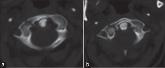
(a and b) Axial view of computed tomographic scan through the atlas demonstrating a right anterior 1/4 isolated fracture with mild distraction (the fracture line goes through the right anterior part of lateral mass)

(a and b) Axial view of computed tomographic scan through the atlas demonstrating a right anterior 1/4 isolated fracture with mild distraction (the fracture line goes through the right anterior part of lateral mass)