
Figure 5.
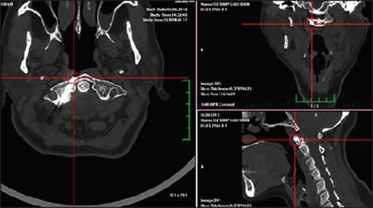
3-dimensional reformatted CT images of the late follow-up images in the axial, sagittal, and coronal planes from thin-section CT scan, demonstrating good reduction with well-positioned screw placement through the right anterior 1/2 single fracture line