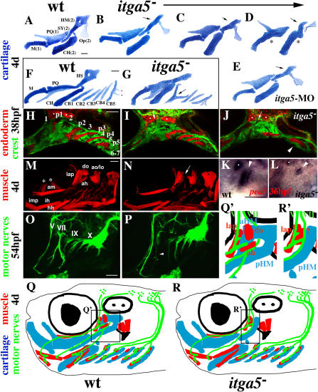
Figure 2. Region-Specific Pharyngeal Defects in integrinα5 Mutants.
(A–E) Flat mount dissections of hyoid and mandibular cartilages from fixed, 4-d-old wild-type (A), integrinα5− (B–D), and itga5-MO (E) animals. Meckel's (M) and palatoquadrate (PQ) cartilages are derived from the mandibular arch (1), and CH, SY, and HM cartilages and the opercle (Op) bone are derived from the hyoid arch (2). A phenotypic series (B–D) shows that the anterior half of HM (arrows) is absent and SY is progressively reduced in integrinα5− animals. Rarely, mandibular and hyoid joints are also missing in integrinα5− animals (asterisks in D). (E) Animals treated with itga5-MO display similar reductions of HM (arrow) and SY.
(F and G) Flat-mount dissections of the pharyngeal cartilages of 4-d-old wild-type (F) and integrinα5− (G) animals. In addition to the mandibular and hyoid cartilages, the five CB cartilages (CB1–CB5) that are derived from the third through seventh arches are shown. Note the teeth on CB5 (dots in F). In integrinα5− embryos we see rare fusions of CB cartilages (arrow in G).
(H–J) Confocal micrographs of the pharyngeal arches of wild-type fli1-GFP (H) and integrinα5−; fli1-GFP (I and J) embryos stained with anti-GFP and Zn8 antibodies at 38 hpf. Neural crest cells of the pharyngeal arches are labeled with fli1-GFP (green, numbered in [H]), and the pharyngeal pouches are labeled by the Zn8 antibody (red, numbered p1–p5 in [H]). In integrinα5−; fli1-GFP embryos, the first pouch is absent or very reduced at 38 hpf (arrows in I and J). Less frequently, we also see reductions in more posterior pouches in integrinα5−; fli1-GFP embryos (arrowhead in J shows a single endodermal mass where p3–p5 would be in wild-type embryos). The Zn8 antibody also recognizes cranial sensory ganglia (dots).
(K and L) In situ hybridizations of wild-type (K) and integrinα5− (L) embryos stained with the pharyngeal pouch marker pea3 at 36 hpf (arrowhead denotes first pouch). The first pouch of integrinα5− embryos is very reduced, but still expresses pea3. Sensory ganglia also stain with pea3 (dots).
(M and N) Cranial muscles of 4-d-old wild-type fli1-GFP (M) and integrinα5−; fli1-GFP (N) embryos stained with MF20 antibody. Mandibular muscles (intermandibularis posterior [imp], adductor mandibulae [am], levator arcus palatine [lap], and do) and hyoid muscles (interhyal , hyohyal [hh], ah, ao, and lo) are labeled in wild-type. integrinα5− embryos have a selective reduction of do and ah muscles (arrow in [N]). Confocal projections of integrinα5− animals did not include ocular muscles (asterisks in M).
(O and P) Cranial motor nerves of wild-type islet1-GFP (O) and integrinα5−; islet1-GFP (P) live embryos at 54 hpf. islet1-GFP-expressing cranial motor neurons innervate muscles of the pharyngeal arches with the following strict segmental correspondence: trigeminal (V)—mandibular; facial (VII)—hyoid; glossopharyngeal (IX)—third; and vagus (X)—fourth through seventh. In integrinα5−; islet1-GFP embryos, facial nerve VII (arrowhead in P) is reduced and/or fails to branch.
(Q and R) Summary of integrinα5 regional pharyngeal defects extrapolated to a 4-d-old embryo and color-coded for cartilage (blue), muscle (red), and nerve (green). Shown in black are the eye (filled circle within larger circle), ear (two dots within oval), and opercle bone (mushroom). In wild-type animals, facial nerve VII innervates and passes by do and ah muscles that are in close association with the aHM cartilage region (enlarged in Q′). In integrinα5 mutants, we see specific reductions of the first pouch (not shown), the aHM cartilage region, do and ah muscles, and facial nerve VII (enlarged in R′). Scale bars: 50 μm.

