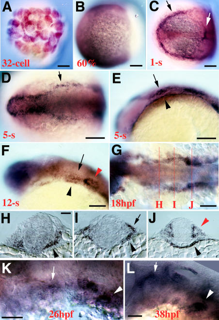
Figure 3. integrinα5 Expression in Pharyngeal Endoderm and Cranial Neural Crest.
(A) At the 32-cell stage, strong maternal integrinα5 expression is seen.
(B) At 60% epiboly, integrinα5 expresses broadly throughout the mesendoderm.
(C) Dorsal view of a 1-s-stage embryo. integrinα5 transcript is concentrated in the ectoderm at the edge of the neural plate (black arrow), in scattered presumptive endodermal cells, and in the first somite (white arrow).
(D and E) Dorsal (D) and lateral (E) views of a 5-s-stage embryo show ectodermal (arrows) and pharyngeal endodermal (arrowhead) expression domains of integrinα5. Ectodermal integrinα5 expression includes migratory hyoid crest, otic placode, and forebrain.
(F) At the 12-s stage, integrinα5 continues to be expressed in the pharyngeal endoderm (black arrowhead), postmigratory hyoid crest (arrow), ear (red arrowhead), and forebrain.
(G–J) At 18 hpf, a dorsal view of an embryo stained for integrinα5 transcript (G) shows approximate axial levels at which cross-sections were prepared. (H) A cross-section at the level of the first pouch shows strong integrinα5 expression in the pharyngeal endoderm (arrowhead). (I) A cross-section at the level of the hyoid arch shows expression of integrinα5 in neural crest (arrow) and pharyngeal endoderm (arrowhead). (J) A cross-section at the level of the ear shows integrinα5 expression in the otic epithelium (red arrowhead) and pharyngeal endoderm (black arrowhead).
(K and L) At 26-hpf (K) and 38-hpf (L) stages, integrinα5 transcript is enriched in the region of the most recent forming pharyngeal pouch (arrowheads) and in patches of crest (arrows).
Scale bars: (A–C), (F), and (G): 100 μm; (D), (E), and (H–L): 50 μm.

