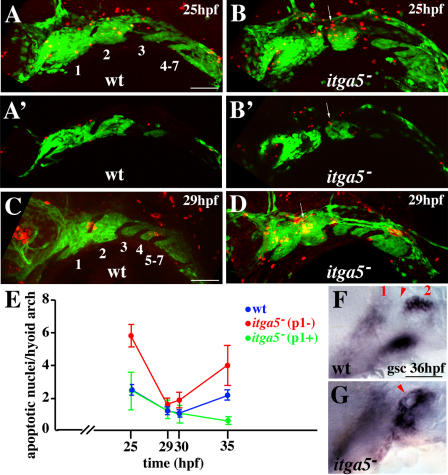
Figure 6. Increased Apoptosis and Disorganized gsc Expression in the Hyoid Arches of integrinα5 Mutants.
(A–D) TUNEL staining of wild-type fli1-GFP (A and C) and integrinα5−; fli1-GFP (B and D) animals shows apoptotic nuclei (red) relative to the GFP-expressing crest of the pharyngeal arches (green) at 25 hpf (A and B) and 29 hpf (C and D). In wild-type confocal projections arches are numbered. (A′) and (B′) are representative confocal sections taken from the projections in A and B. In integrinα5− animals lacking the first pouch, increased apoptosis (arrows in [B] and [D]) is seen in the dorsal anterior hyoid arch adjacent to where the first pouch would be in wild-type animals. In mutant sections (B′), TUNEL-positive cells (arrow) colocalize with the fli1-GFP crest marker.
(E) The number of apoptotic nuclei per hyoid arch is plotted versus time for wild-type sides (blue) and integrinα5− sides without (p1−; red) or with (p1+; green) a normal first pouch. At 25 hpf, integrinα5− hyoid arches had more apoptotic nuclei than wild-type hyoid arches only when the first pouch was defective (p < 0.0001). At later time points, integrinα5− hyoid arches missing the first pouch had a tendency to have more apoptotic nuclei than wild-type or integrinα5− arches with normal first pouches (only itga5− with a normal first pouch versus itga5− without at 35 hpf is statistically significant, p < 0.05). Total sides examined: 25 hpf: nwt = 40, nitga5 = 38; 29 hpf: nwt = 30, nitga5 = 26; 30 hpf: nwt = 30, nitga5 = 20; and 35 hpf: nwt = 30, nitga5 = 14.
(F and G) gsc expression at 36 hpf labels dorsal and ventral domains of hyoid crest. Mandibular (1) and hyoid (2) arches are numbered, and the first pouch is denoted by arrowhead. In wild-type animals, dorsal and ventral hyoid gsc domains are well separated. In this integrinα5− animal, dorsal and ventral hyoid gsc domains are fused, and disorganized gsc-expressing cells envelop the reduced first pouch (arrowhead).
Scale bars: 50 μm.

