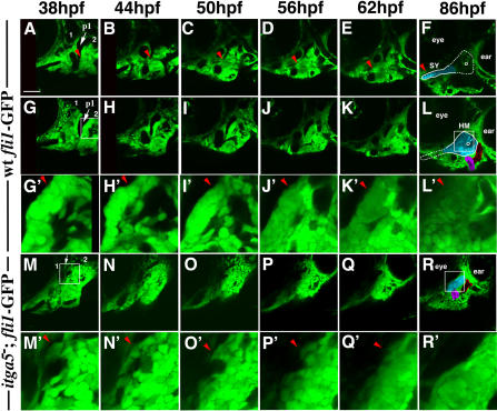
Figure 7. Anterior Hyoid Crest Cells Display Aberrant Behavior in integrinα5 Mutants.
Confocal time-lapse recordings show hyoid cartilage development in wild-type fli1-GFP (Videos S1 and S2) and integrinα5−; fli1-GFP (Video S3) animals from 38 hpf to 86 hpf (nwt = 3; nitga5 = 4). Videos S1 and S2 are different depths of the same time-lapse recording. Representative imaging stills of Video S1 (A–F), Video S2 (G–L), and Video S3 (M–R) were taken at 38 hpf (A, G, and M), 44 hpf (B, H, and N), 50 hpf (C, I , and O), 56 hpf (D, J, and P), 62 hpf (E, K, and Q), and 86 hpf (F, L, and R). At the beginning of the recordings (A, G, and M), the mandibular (1) and hyoid (2) arches are numbered and an arrow denotes the first pouch (p1). At the end of the recordings (F, L, and R), the cartilage regions are clearly visible as large cells with thick matrix (pseudocolored blue). The outline of the HS cartilage, a composite of SY and HM regions, is shown in (F) and (L). As a reference, the opercle bone and ao/lo hyoid muscle mass are pseudocolored purple and red, respectively, and the eye and ear are labeled. In Video S1 (A–F), red arrowheads denote a cluster of cells adjacent to the first pouch that undergo cellular rearrangements and form the long, anterior SY extension in wild-type animals. (G′–R′) show magnifications of HM-forming regions taken from (G–R) and correspond to areas within white boxes given in (G) and (L) for (G–L) and in (M) and (R) for (M–R). In wild-type development, hyoid crest cells adjacent to the first pouch remain a tightly packed mass as aHM chondrifies (e.g., cells denoted by red arrowheads in G′–L′). In integrinα5 mutants, the first pouch is missing (white arrow in [M]), and anterior hyoid crest cells are disorganized at 38 hpf (e.g., arrowhead in [M′]). Over time, anterior hyoid crest cells migrate out of the region and do not contribute to cartilage (e.g., arrowheads in [N′–Q′]). In contrast, the pHM region and the opercle bone develop normally from more posterior hyoid crest in integrinα5− animals (R). Scale bar: 50 μm.

