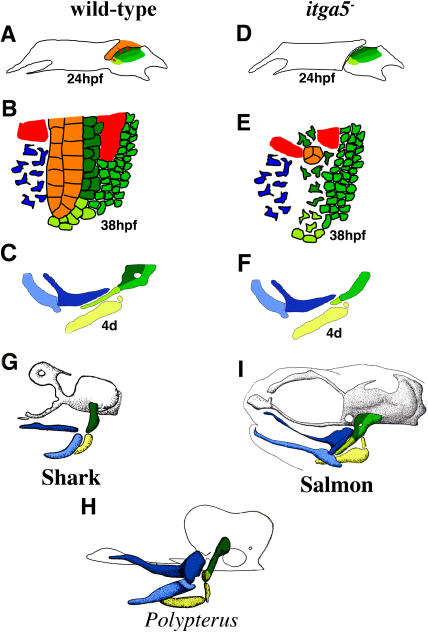
Figure 8. Model for Development and Evolution of Hyoid Cartilage.
(A–F) Models of hyoid development in wild-type (A–C) and integrinα5− (D–F) animals show the structure of hyoid arches at 24 hpf (A and D) and 38 hpf (B and E) and mandibular and hyoid cartilages at 4 d (C and F).
(A) At 24 hpf of wild-type development, crest that will form aHM (dark green), pHM (medium green), and SY (light green) cartilage regions occupy distinct domains within the hyoid arch. Signals (red arrows) from the first pouch (orange) stabilize adjacent aHM- and SY-producing crest.
(B) At 38 hpf of wild-type development, aHM- and SY-producing crest tightly pack along the first pouch. Cranial mesoderm (red) and some mandibular crest (blue) are also shown.
(C) At 4 d of wild-type development, the HS cartilage is a composite of aHM, pHM, and SY regions. Also shown are the hyoid CH (yellow) and mandibular Meckel's (light blue) and palatoquadrate (dark blue) cartilages.
(D) In integrinα5− animals, the first pouch is missing or very reduced at 24 hpf.
(E) By 38 hpf, as a consequence of the lack of a first pouch, aHM and SY progenitors are disorganized and undergo gradual apoptosis. In contrast, the development of pHM progenitor cells does not require the first pouch.
(F) At 4 d, aHM and SY cartilage regions are selectively reduced in integrinα5− animals.
(G–I) The HS element has undergone extensive change during vertebrate evolution. In the illustrations (adapted from De Beer [1937]), the neurocranium is grey or outlined in black and mandibular and hyoid cartilages are color-coded as described above. Based on relations to morphological landmarks and data presented here on the tripartite mosaic development of HS, an evolutionary scheme is proposed.
(G) In the dogfish shark Scyliorhinus canicula, a single rod-shaped element corresponds to pro-aHM/SY regions.
(H) In the basal actinopterygian Polypterus senegalus, separate aHM and SY regions are present.
(I) As shown for salmon, during actinopterygian evolution a new region, pHM, develops posterior to and fuses with aHM to create a wide HM plate that articulates with the neurocranium and supports an enlarged, overlying opercular apparatus (not shown).

