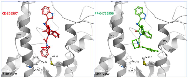
Figure 8. Comparison of docked binding poses of CE-326597 and PF-04756956 at CCK1R.
Shown are the best performing models illustrating the docking pose of CE-326597 (left) and PF-04756956 (right) from a side view. The binding poses of both compounds were predicted to be accommodated in a similar pocket, however the differences between the two are marked by the position of the N1-benzyl (CE-326597) that is buried deeper, and the dimethyl benzyl at C2 position of piperidine (PF-04756956) that is more superficially located. Also shown are potential interacting residues, Trp 6.48, Met 3.32 and Val 3.36. Large contact areas between the side-chain and ligand are represented by thicker stick representation on the mutated residues.

