
Figure 12.
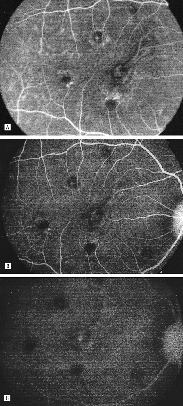
Fluorescein angiogram of one eye after intravenous injection with fluoresceinated anti-VEGF antibody, demonstrating localization to experimental choroidal neovascularization. Hyperfluorescence noted 1 minute after injection (A), 20 minutes after injection (B), and with leakage noted one hour after injection. Reproduced with permission from Tolentino MJ, Husain D, Theodosiadis P, et al. Angiography of fluoresceinated anti-vascular endothelial growth factor antibody and dextrans in experimental choroidal neovascularization. Arch Ophthalmol. 2000;118:78–84. Copyright 2000 American Medical Association.27