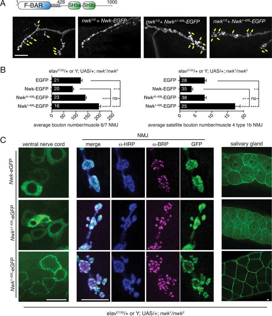
Figure 1. The Nwk F-BAR domain is required for its in vivo function.
(A) Schematic of Nwk domain structure and representative confocal images of α-Cpx-stained third instar larval NMJ morphology on muscle 4 for the indicated genotypes. Arrows indicate satellite boutons. (B) Quantification of synaptic growth on muscle 6/7 and satellite bouton number on muscle 4 by Nwk variants in a nwk1/nwk2 null background. Graphs show mean +/− s.e.m. Numbers in bar graphs represent the number of NMJs. (C) Localization of Nwk variants. GFP-tagged Nwk variants were expressed in the nwk1/nwk2 null background, under control of the GAL4 driver elavC155 (pan-neuronal and salivary glands). Third instar larvae were fixed and stained with α-BRP and α-HRP antibodies. Scale bars are 10 µm. Associated with Fig. S1.

