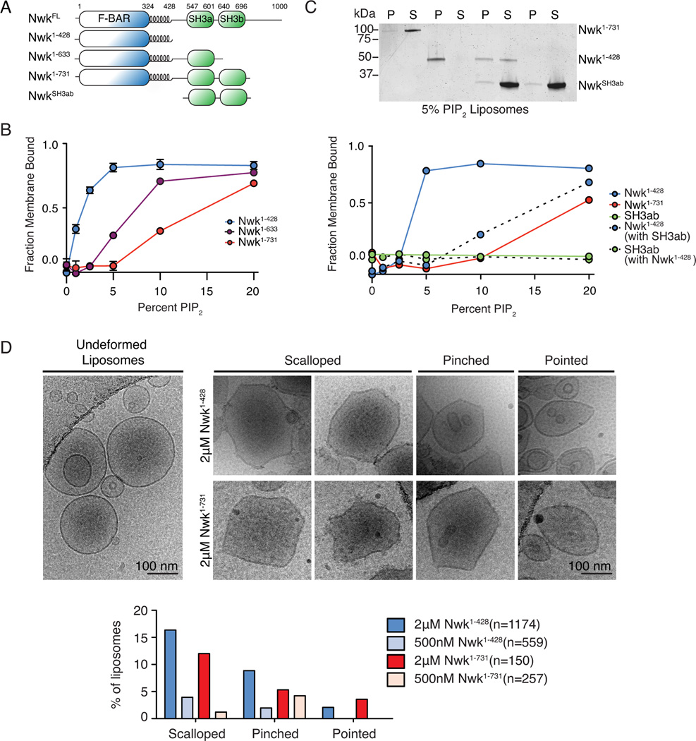
Figure 4. Nwk SH3b inhibits membrane deformation by the Nwk F-BAR domain.
(A) Nwk constructs used for in vitro assays. (B, C) Purified proteins (10 µM NwkSH3ab (Nwk residues 536–731); 1 µM all other proteins) were incubated with liposomes of the following composition: 80-X% PC, 15% PE, 5% PS and X% PI(4,5)P2 (where X is the concentration indicated in the graph) and subjected to liposome cosedimentation assays. Graphs show mean densitometry from one (B) or three (mean +/− s.e.m.) (C) independent experiments. (C) Image shows representative Coomassie staining of supernatant (S) and pellet (P) fractions at 10% PI(4,5)P2. (D) Cryo-EM of control and Nwk deformed liposomes. Both purified Nwk1–428 and Nwk1–731 induce membrane scalloping, pointing, and pinching of 10% PI(4,5)P2 liposomes. Scale bar is 100 nm. Bar graph summarizes vesicle morphology after 30-minute incubation of Nwk1–428 or Nwk1–731 (2µM and 500nM) with 0.3 mM [DOPC:DOPE:DOPS:PI(4,5)P2] = 70:15:5:10 liposomes. n represents the number of liposomes examined; Associated with Fig. S3.

