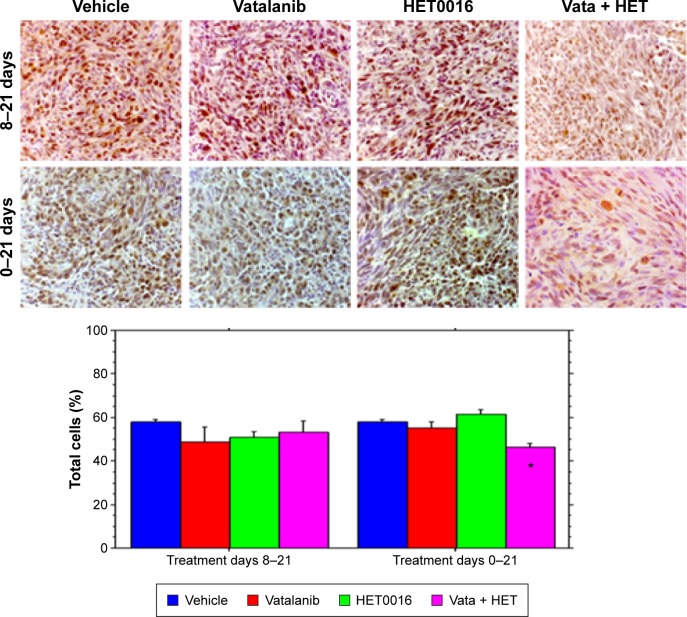
Figure 6.
Proliferative capacity.
Notes: Histochemical images and analysis of proliferative capacity of the tumor cells following different treatments. Five different regions with higher expression of ki67 were taken within the tumor mass, and brown and blue cells were counted in total. The total number of browns cells (Ki-67 stained) was divided by the total number of cells (blue and brown cells). Combined HET0016 and vatalanib treatment started on day 0 caused significantly lower proliferative cells in the tumor. IHC images were taken at ×40 magnification. *Significant difference from vehicle-treated animals (P<0.05).
Abbreviations: HET0016, N-hydroxy-N′-(4-butyl-2-methylphenyl)formamidine; IHC, immunohistochemistry; Vata, vatalanib.

