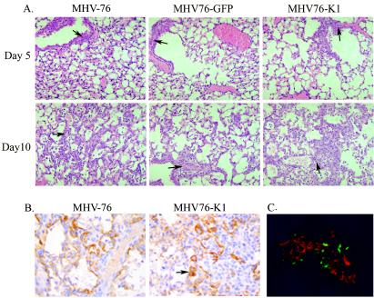
FIG. 5.
(A) Hematoxylin and eosin staining of lung sections of mice infected with MHV-76, MHV76-GFP, or MHV76-K1 at days 5 and 10 p.i., showing perivascular inflammatory cell infiltrates (arrows). (B) Cytokeratin staining of lung sections of mice infected with MHV-76 or MHV76-K1 at day 10 p.i., showing strong staining of type 2 pneumocytes in chronic proliferative inflammatory foci (arrow). (C) Mice infected with MHV76-K1 express GFP in the lungs at day 5 p.i. MHV-76 antigens were stained with polyclonal antisera and an anti-rabbit tetramethyl rhodamine isocyanate conjugate (red).

