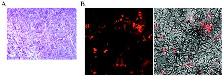
FIG. 6.
(A) Hematoxylin and eosin staining of the salivary gland adenocarcinoma present in an MHV76-K1-infected mouse at day 120 p.i. (B) Expression of MHV-76 antigens in the salivary gland adenocarcinoma detected with rabbit polyclonal antisera. The left panel shows the immunofluorescent image, and the right panel shows a phase-contrast image merged with the immunofluorescent image.

