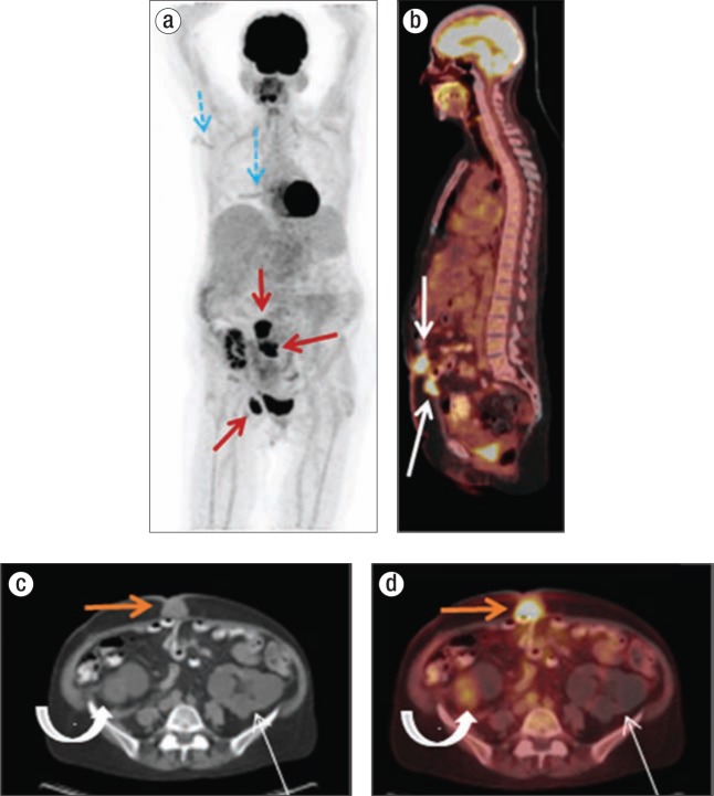
Figure 2.
(a) Whole-body MIP image from an F-18 FDG PET/CT demonstrates abnormal radiotracer uptake within the two separate regions of the anterior abdominal wall as well as the right inguinal region (solid red arrows). Linear uptake in the right back and axilla (blue dashed arrows) corresponds to the melanoma excision and axillary node dissection sites. (b) Sagittal whole-body image from the same patient shows abnormal focal radiotracer uptake in two separate areas of the anterior abdominal wall (white arrows). (c) Transaxial CT and (d) FDG-18 PET/CT images at the level of the superiorly located abdominal wall mass show abnormal radiotracer uptake in the midline anterior wall on FDG-18 PET/CT, which corresponds with the anterior abdominal wall mass seen on CT (orange arrow). Also shown in these images are a right renal transplant (curved arrow) and polycystic left kidney (thin arrow).

