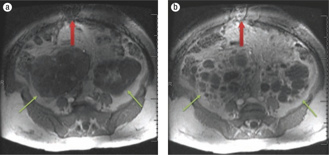
Figure 3.
(a) Precontrast and (b) postcontrast transaxial spoiled gradient echo MR images from November 2005, which depict an enhancing anterior periumbilical mass that was biopsied in September 2014 and proven to be epithelial-type peritoneal mesothelioma (red arrow). Changes of polycystic kidney disease are also shown in these images (green arrows).

